|
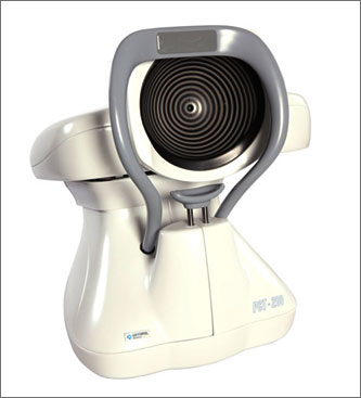
Thanks to advanced digital image
analysis methods a new standard in
corneal topography has been developed. |
|
The device is fully automatic. There is
no need to manually align position of
the bowl. All this is done by the
computer. |
|
The advantages are simple: faster,
better and more precise measurement. |
|
|
|
|
|
TECHNICAL DATA |
|
|
|
height: 515 mm |
|
width: 260 mm |
|
depth: 355 mm |
|
projection method: placido rings
projection |
|
pattern colour: pure red |
|
measurement method: digital image
analysis |
|
number of rings: 19 |
|
number of analysed points: 16000 |
|
papping methods: |
|
|
|
local curvature tangential map, |
|
sagital map, |
|
results comparison, |
|
height map, |
|
3D map |
|
|
|
display units: refractive power (diopters)
curvature (mm) |
|
utilities: full keratometric data, map
distance, |
|
cross-section measurement |
|
power supply: 230V/50Hz |
|
|
|
MINIMUM COMPUTER REQUIREMENTS |
|
|
|
operating system: Win 2000/XP |
|
processor: Pentium III 1 GHz |
|
RAM memory: 128MB |
|
HDD 40 GB |
|
video: 800x600 screen resolution in True
Colour |
|
communication port: USB 2.0 (HI-SPEED) |
|
|
|
OTHER REQUIREMENTS |
|
|
|
printer: colour printer required |
|
recommended HP DeskJet series printer. |
|
|
|
SOFTWARE FEATURES |
|
|
|
Fourier analysis window |
|
Zernike analysis window |
|
possibility of mutual movement of map
and pattern in height window |
|
possibility to store several fluorescent
stimulations |
|
very low bowl illumination during bowl
alignment |
|
very short flash during examination
(250ms) |
|
internal database |
|
automatic data exchange with PTS series
perimeters |
|
new improved software for fitting hard
contact lenses |
|
fluorescent stimulation of hard contact
lens fitting |
|
possibility of working in computer net
simultaneously by several users |
|
easier configuration process (auto-config
function available, setting
automatically instrument parameters) |
|
possibility of changing the degree of
transparency of displayed map in
relation to an eye in the background |
|
possibility of manual edition of
position and size of detected iris and
pupil |
|
possibility of manual edition of
detected rings |
|
print preview of examination results
with possibility of saving to graphic
file |
|
automatic calibration module |
|
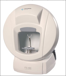
Thanks to advanced digital image
analysis methods a new standard in
corneal topography has been developed. |
|
The device is fully automatic. There is
no need to manually align position of
the bowl. All this is done by the
computer. |
|
The advantages are simple: faster,
better and more precise measurement. |
|
|
|
|
|
TECHNICAL DATA |
|
|
|
height: 515 mm |
|
width: 260 mm |
|
depth: 355 mm |
|
projection method: placido rings
projection |
|
pattern colour: pure red |
|
measurement method: digital image
analysis |
|
number of rings: 19 |
|
number of analysed points: 16000 |
|
papping methods: |
|
|
|
local curvature tangential map, |
|
sagital map, |
|
results comparison, |
|
height map, |
|
3D map |
|
|
|
display units: refractive power (diopters)
curvature (mm) |
|
utilities: full keratometric data, map
distance, |
|
cross-section measurement |
|
power supply: 230V/50Hz |
|
|
|
MINIMUM COMPUTER REQUIREMENTS |
|
|
|
operating system: Win 2000/XP |
|
processor: Pentium III 1 GHz |
|
RAM memory: 128MB |
|
HDD 40 GB |
|
video: 800x600 screen resolution in True
Colour |
|
communication port: USB 2.0 (HI-SPEED) |
|
|
|
OTHER REQUIREMENTS |
|
|
|
printer: colour printer required |
|
recommended HP DeskJet series printer. |
|
|
|
SOFTWARE FEATURES |
|
|
|
Fourier analysis window |
|
Zernike analysis window |
|
possibility of mutual movement of map
and pattern in height window |
|
possibility to store several fluorescent
stimulations |
|
very low bowl illumination during bowl
alignment |
|
very short flash during examination
(250ms) |
|
internal database |
|
automatic data exchange with PTS series
perimeters |
|
new improved software for fitting hard
contact lenses |
|
fluorescent stimulation of hard contact
lens fitting |
|
possibility of working in computer net
simultaneously by several users |
|
easier configuration process (auto-config
function available, setting
automatically instrument parameters) |
|
possibility of changing the degree of
transparency of displayed map in
relation to an eye in the background |
|
possibility of manual edition of
position and size of detected iris and
pupil |
|
possibility of manual edition of
detected rings |
|
print preview of examination results
with possibility of saving to graphic
file |
|
automatic calibration module |